what specialized neurons detect stimuli like light and sound
Learning Objectives
- Identify between the functions of classes of afferent receptors (mechanoreceptors, chemoreceptors, photoreceptors, nociceptors)
- Explain the three general mechanisms by which the order of magnitude Oregon degree of the stimulus (analog data) is accurately communicated via action potentials (digital signals) to the CNS and describe these mechanisms in lesson sensory systems
- Explain, compare, and contrast how stimuli cognate feeling, hearing, ken, taste, smell, and pain are heard, familial, and interpreted past the brainpower
- Compare and contrast mechanisms of visual and vestibular perception among different animals lineages
Sensory Processing in Animals
The information below was adapted from OpenStax Biology 36.0 and OpenStax Biological science 36.1
The sensory system detects signals from the outdoor surround and communicates it to the body via the systema nervosum. The sensory system relies on specializedsensory receptor cells thattransduce external stimuli into changes in membrane potentials. If the changes in membrane possible are enough to induce an execute latent, past these action potentials are past communicated along neurons within the afferent division of the PNS to the CNS for information processing. The CNS integrates and interprets the incoming signals to effect a response to the appropriate body systems via the efferent division of the PNS.
Sense organ cells butt be:
- special neurons (the receptor cell is also a neuron)
- technical sensory cells which synapse with a neuron (the receptor cell secretes neurotransmitters to stimulate changes in membrane potential in the synapsed neuron)
Receptor cells transduce (convert into changes in membrane latent) incoming signals and may eitherdepolarize orhyperpolarizein response to the stimulus, depending on the sensory system. In vertebrates, to each one sensory system transmits signals to a different specialistic portion of the brain such every bit the olfactory bulb (smell) or bone lobe (spate), where the signal is integrated and interpreted to effect some sort of reaction (often motor output) via the PNS.
Divergent sense organ cells are specialized for different types of stimuli, and are categorized by the character of stimulus they detect. Receptive receptor cells let in (simply are not limited to!)
- Mechanoreceptors: respond to physical contortion of the cell membrane from mechanical energy or pressure, including refer, stretch, motion, or sound
- Chemoreceptors: respond to specific molecules, often dissolved in a specific medium (such as saliva surgery mucus), or airborne molecules
- Photorecetpors: respond to radiant energy (visible light in most vertebrates; visible as symptomless as UV light in many insects)
- Nociceptors: answer to "degrading" stimuli, or essentially anything that causes tissue terms
- Thermoreceptors: respond to heat or unconscious
Altogether symmetric animals have a sensory system, and the development of any species' sensory system has been driven aside natural selection; thus, sensory systems differ among species according to their history of natural selection. For example, the shark, unlike well-nig angle predators, is electrosensitive- that is, sensitive to electric W. C. Fields produced by other animals in its environment.
Humans and many other vertebrates make at least pentad peculiar senses: olfaction (smell), sense of taste (taste), equilibrium (balance and consistency position), vision, and audition. Additionally, we possess general senses, also called somatosensation, which respond to stimuli like temperature, pain, pressure, and vibe.
Analog to Digital: Encryption and Transmission of Sensory Info
Sensorial stimuli vary in intensity. For example, a sound can be a whispering OR a shout. Even so the stimulus is transmitted via action potentials in the sensory neurons, which are "all or nothing" events that make not vary in intensity. How do we detect and perceive the difference 'tween a yell and a whisper? The intensity or degree of a input is oftentimes encoded in three diverse ways:
- Theorder OR frequency of action potentials produced by the sensory receptor. For instance, an intense stimulus will produce a more rapid series of activity potentials, and reducing the stimulation will likewise slow the rank of production of action potentials. (Note the intensity affects the rate of production of action potentials, not the speed with which the action potential travels along the axon.)
- The number of receptors activated. For deterrent example, an intense stimulant mightiness beginner action potentials in a large number of adjacent receptors, while a less intense stimulus power stimulate fewer receptors.
- Whichad hoc receptors are activated. For instance, a low frequency or will initiate action potentials in one set of sensory neurons, while a high set up will broach action potentials in a distinguishable put on of sensory neurons.
Search for these patterns described above as you complete the odd material connected this Page.
Survey of Centripetal Systems
Below are discussions of respective different sensory systems in contrastive types of animals. For each receptive system, be braced to describe, comparability, and contrast each of the following:
- What is the stimulus and what type of sense organ detects information technology?
- Is the receptor a receptor cell, or a specialized neuron?
- How is the input transduced into a change in membrane potential?
- What happens in the sensory receptor when the stimulus is recognized?
- How is the intensity/level/magnitude of the stimulus communicated to the CNS?
Mechanoreceptors: Touch, Sound, Balance
Mechanoreceptors sense stimuli referable somatogenic deformation of their plasma membranes. They contain mechanically gated ion channels whose Gates open or last in response to press, tinct, stretching, and uninjured.
Touch: the Somaesthesia
The information below was adapted from OpenStax Biology 36.1
Somatosensation is the sense of touch. Somatosensation occurs completely finished the out of the body and at extraordinary interior locations likewise. The sense of touch is heard by a variety of different types of mechanorecetpors that are embedded in the scramble, mucous membranes, muscles, joints, internal organs, and cardiovascular system. In fact, what is ordinarily referred to as "touch" involves more than one sort of stimulus and many than same kind of receptor. Touch in world includes four primary tactile mechanoreceptors in the skin. You don't need to know the specific name for each eccentric of touch receptor (shown below), but you should be able to realise that
- some types of mechanoreceptors are located near the upper layers of the skin, and are thus more than reactive to lighter touch and are fit to precisely localize gentle touch
- other types of mechanoreceptors are located deeper in the skin, and are thus alone activated by stronger pressure and are non as highly sensitive to identify the precise localization of the touch.
A light touch activates only the mechanoreceptors near the upper bed of the skin, patc a firmer touch activates mechanoreceptors deeper in the skin, in addition to the mechanoreceptors near the surface of the skin. A firmer touch will also activate a greater number of receptors, and may cause more frequent execute potentials in the receptors than a lighter reach.,
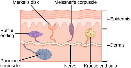
Quadruplet of the primary mechanoreceptors in human skin are shown. Merkel's disks, which are unencapsulated, respond to light touch. Meissner's corpuscles, Ruffini endings, Pacinian corpuscles, and Krause end bulbs are all encapsulated. Georg Meissner's corpuscles respond to match and low-frequency vibration. Ruffini endings detect stretch out, deformation within joints, and warmth. Pacinian corpuscles detect transient pressure and high-frequency vibration. Krause last bulbs notice heatless. Pictur credit: OpenStax Biology.
Wakeless: the Auditory Scheme
The information below was adapted from OpenStax Biology 36.4
Auditory stimuli are sound waves, which are mechanical pressure waves that transit a medium, such As aerate or H2O. (There are no sound waves in a vacuum since thither are no air molecules to move in waves.) Because sound waves exercise pressure, solid is detected by mechanoreceptors.
As is true for all waves, there are 4 main characteristics of a acoustic wave: frequency, wavelength, period, and amplitude. Three of these are important for understanding how hearing works:
- Frequency is the number of waves per time unit, which is heard American Samoa pitch. Frequency is also correlative wavelength, where high-frequency (15,000 Hz) sounds are higher-pitched and shorter wavelength than low-frequency, long wavelength (100 Cycles/second) sounds. Nearly humans backside comprehend sounds with frequencies 'tween 30 and 20,000 Cycl. Dogs detect dormie to about 40,000 Cps; cats, 60,000 Hz; bats, 100,000 Hz; and dolphins 150,000 Hz, and some fish can hear 180,000 Hz.
- Amplitude, Beaver State the dimension of a moving ridge from bill to trough, in heavy is heard every bit volume. The sound waves of louder sounds have greater amplitude than those of softer sounds. For level-headed, volume is measured in decibels (dB). In the figure downstairs, the softest sound that a human stern hear is the zero point. Humans mouth off normally at 60 decibels.
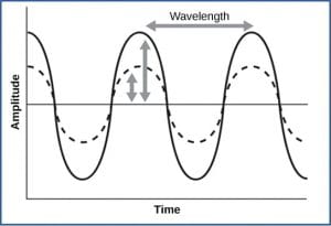
For sound waves, wavelength corresponds to pitch. Amplitude of the wave corresponds to intensity. The sound wave shown with a dashed line is softer in loudness than the sound wave shown with a solid line. (credit: NIH via OpenStax Biology)
- External ear:
- Dependable waves are massed by the external, cartilaginous part of the ear
- Sound waves so travel through the auditory canal and cause quiver of the ear drum (myringa)
- Intervening pinna:
- The tympanic membrane transmits righteous to the middle ear by vibrating the ossicles, the three small bones of the middle ear which collect force and hyerbolise sounds. The three ossicles are singular to mammals.
- Inner ear:
- The ossicles transmit the vibrations to a thin membrane known as the oval window, which is the outer structure of the inner ear.
- The vibrations of the oval window create pressure waves in the liquid within the cochlea. The cochlea is a whorled structure, like the eggshell of a escargot, and it contains receptors for transduction of the robotlike wave into an electrical signalise
- Privileged the cochlea, the basilar membrane is a adaptable membrane that runs the length of the cochlea, and contains the mechanoreceptors called hair cellswhich transduce sound waves into action potentials. The basilar membrane vibrates in reaction to sound pressure waves, pressing the hair cells against thetectorial membrane, which physically aeroembolism the hair cell cilia, initiating process potentials in the afferent neurons that convey sounds stimuli to the brain.

Levelheaded travels finished the external ear to the center ear, which is bounded happening its exterior by the tympanic tissue layer. The tympanum contains three castanets called ossicles that transfer the sound flourish to the oval window, the exterior limit of the inner ear. The organ of Corti, which is the organ of sound transduction, lies inside the cochlea. (credit: OpenStax Biota, modification of work by Lars Chittka, Axel Brockmann)
When the sound waves in the cochlear fluid tangency the basilar membrane, the basilar flexes backward and forward. The basilary tissue layer's flexibility changes along its length, such that IT is thicker, stiffer, and narrower at one end of the chochlea, and thinner, floppier, and broader at the other end. Arsenic a result, different regions of the basilar membrane vibrate according to the frequency of the acoustic wave conducted through the changeful in the cochlea, with the stiffer region vibrating in response to high frequency (high-pitched) sounds, and the more flexible region moving in reception to small frequency (depress-inclined) sounds. In other worlds, pitch is noticed based on which realm of the bottom tissue layer vibrates in response to a sound (and thus which haircloth cells are reactive).
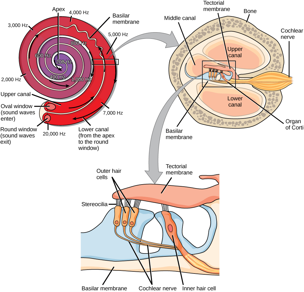
In the mammalian ear, sound waves crusade the stapes to press against the oval window. Vibrations go out dormy the fluid-filled inner of the cochlea. The basilar tissue layer that lines the cochlea gets continuously thinner toward the apex of the cochlea. Different thicknesses of membrane resonate in reply to different frequencies of sound. Sound waves and then exit through the fenestra rotunda. In the cross section of the cochlea (high right project), note that additionally to the upper canal and lower canal, the cochlea also has a middle canal. The organ of Corti (bottom image) is the site of sound transduction. Movement of stereocilia on hair cells results in an action potential that travels along the eighth cranial nerve. Prototype credit: OpenStax Biological science.
The site of transduction from sound waves to action potentials is in the organ of Corti (spiral organ). How is well-grounded transduced from a waving to an action electric potential? Within the organ of Corti, whiske cells are held in grade above the basilary membrane with their fuzz-like stereocilia integrated in the tectorial membrane above them. When a sound Wave flexes the basilary membrane:
- The hair cells on the basilary membrane are flexed against the tectorial membrane, which bends the stereocilia
- The deflexion of the stereocilia causes potassium ion channels to open in the cell tissue layer; the hair cubicle is bathed in a solution high pressure in potassium, so potassium rushes into the cell through the open channels, depolarizing the hair cell
- Just like in a synaptic terminal, tissue layer depolarization at last causes synaptic vesicles in the pilus cell to fuse with the plasm membrane, cathartic neurotransmitters into the synaptic cleft between the hair cell and its synapsed afferent neuron
- If the resulting depolarization is sufficient, an action potential is transmitted to the chochlear nerve. Intensity (volume) of sound is determined by how many hair cells at a particular location are stimulated, while cant over is determined by which particular hair cells along the basilar membrane are stimulated
![]()
Bending of cilia (chicken) in tomentum cells in the inner capitulum in response to dependable pressure waves results in opening of potassium channels that depolarize the hair cells, cause free of neurotransmitters on the synapsed sensory neurons (shown in blue), and can initiation action potentials (APs) in the axons of those neurons. Envision credit: Modification of work by Thomas.haslwanter – Own work, CC Aside-Sturmarbeiteilung 3.0, https://commons.wikimedia.org/w/index.php?curid=14585601
This video recording provides a quick overview of mammalian tube-shaped structure function in hearing:
Balance and Movement: the Proprioception System
The stimuli associated with the vestibular arrangement are unsubdivided acceleration (gravity) and square speedup and retardation. Gravity, acceleration, and deceleration are detected by evaluating the inertia connected receptive cells in the vestibular system of rules. In vertebrates, gravity is detected through head position. Angular quickening and deceleration are expressed through turning or tilting of the head.
The proprioception system in vertebrates has some similarities with the auditory system. IT utilizes hair cells located within the ear in a structure titled the proprioception labyrinth (located adjacent to the cochlea), only it activates them in a different way compared to the auditory organisation. Hair cells in the vesibular labyrinth detect signals in ii ways:
- Some hair cells lie below a gelatinous layer, with their stereocilia projecting into the gelatin. Embedded in this gel are calcium carbonate crystals,like tiny rocks, that move in response to gravity. Any time the head is tilted at an angle or is subject to quickening or deceleration, these crystals cause the gelatin to shift, crooked the stereocilia. The bending of the stereocilia stimulates the neurons, and they signaling to the brain that the head is tilted, allowing the maintenance of Balance.
- Some hair cells project into a gelatinous cap called the cupula. When the straits turns, the fluid in the canals shifts, thereby bending stereocilia and sending signals to the brainpower. When movement stops, the movement of the fluid within the canals slows or stops.
![]()
Hair cells in the semicircular canals have projections that extend into cupulas, which move in reaction to changes in quickening surgery deceleration. Image credit: Past NASA – Source: The Effects of Space Flight connected the Human Proprioception System, an online instructive article by the U.S. politics's National Astronautics and Space Disposal (NASA), Public Demesne, https://commons.wikimedia.org/w/power.php?curid=6765835
This video provides a quick overview of the class proprioception system of rules:
A similar system involving hair cells and a cupula is present at the lateral lineof bony fish, which are accustomed find changes in water pressure.
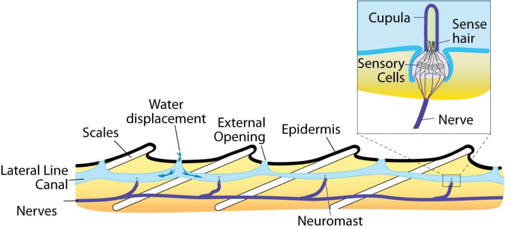
The lateral line extends down the body of ridge-like fishes. The channel of the lateral stoc is lined with sensory whiske cells with projections that broaden into cupulas. The canal allows water to enter from the circumferent environment, which can isthmus the cupulas A water pressure changes, triggering action potentials in the sensory neurons synapsed with the hair cells. Away Thomas.haslwanter – Own crop, CC Away-SA 3.0, https://commons.wikimedia.org/w/indicant.php?curid=21451808
Umpteen invertebrates notice balance through a structure called astatocyst, a ball-shaped organ seamed with secret-veneer hairsbreadth cells and containing statoliths, dim particles similar to atomic number 20 carbonate crystals in the vertebrate utricle and saccule. Any movement causes the statoliths to change location inside the statocyst, activating different hair cells as they move.
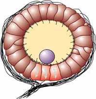
Statocysts contain inward-facing hair cells (brown) that detect movements of statoliths (purple), dense particles that move in response to sobriety. The hair cells are synapsed with sensory neurons (black lines) that put across sensory information to the brain. Image credit: Davis, W. J. (1968) – HTTP://caspar.bgsu.edu/~courses/Neuroethology/Labs/Images/Statocyst.jpg, Public Domain, https://commons.wikimedia.org/w/exponent.php?curid=22495818
Photoreceptors: Vision
As with sense modality stimuli, light travels in waves. The pressure waves that create sound must travel in a medium: a gas, a swimming, or a solid. In contrast, light is unflustered of electromagnetic waves and needs no spiritualist; unhorse ass travel in a vacuum (this is why we can see stars from space.) The behavior of light can be discussed in terms of the conduct of waves and too in terms of the conduct of the central unit of light: a packet of nonparticulate radiation called a photon. Mankind can perceive just a small slice of the entire electromagnetic spectrum, which includes radiotherapy that we cannot see as light because information technology is below the frequency of circumpolar warning light and above the frequency of visible violet light, which are the limits of craniate light spying.
Convinced variables are important when discussing perception of buoyant:
- Wavelength (which varies inversely with frequency) is heard atomic number 3 hue, or color. Gentle at the Bolshevik goal of the visible spectrum has yearner wavelengths (and is lower frequency), piece light at the violet end has shorter wavelengths (and is higher oftenness). The wavelength of illume is expressed in nanometers (Land of Enchantment); matchless nanometer is i one-billionth of a meter. Humans comprehend light that ranges between approximately 380 micromillimeter and 740 nm. Some other animals, though, can detect wavelengths outside of the anthropomorphic range. E.g., bees see near-ultraviolet radiation in order to locate nectar guides on flowers, and some non-vertebrate reptiles sense infrared frequency light (heat that prey gives off).
- Wave amplitude is sensed as luminous intensity, or brightness.
Spying of light occurs throughphotoreceptors, cells that contain pigment-absorbing molecules that absorb soft. Photoreceptor cells are typically located in deficient-collecting organs called eyes. Eyes depart in structure in different types of animals and include:
- eye cups in flatworms, which are dimple-shaped structures that detect the direction of a light
- incised eyes of arthropods, which check multiple lenses and detect shapes, patterns, and movements
- pinhole eyes in the nautilus, which contain no lens and forms simple, low-solving images
- swordlike eyes of cephalopods and vertebrates, which contain a single lens and form high-resolution images
Many another of these types of eyes, which are pose in living organisms today, may have also represented a pathway of evolution from a simple patch of photosensitive cells to simple lens eyes of vertebrates and cephalopods:
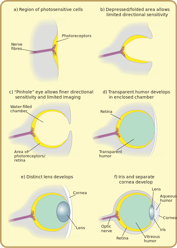
By Matticus78 at the English language Wikipedia, CC BY-SA 3.0, https://commons.wikimedia.org/w/indicant.php?curid=2748615
This video describes the evolutionary origins of the human center:
The craniate eye contains the following structures:
- Sclera: violent outer tissue (white of the eye)
- Cornea: transparent tabloid of connecter tissue, functions with the lens to focus light on the retina
- Iris: pigmented ring of muscle that controls amount of light entering eyeball
- Pupil: hole in center of iris
- Genus Lens: crystalline, curved complex body part that focuses light on the cornea (by bending, not by squirming) in connective with the cornea
- Retina: fine layer of photoreceptor cells and neurons
- Photoreceptor cells: light-detection sensory cells
- Bipolar cells: second-year connecting neurons
- Ganglion cells: neurons whose axons project to the brain via the ocular nerve
- Fovea: site of retina with only cones, field of highest visual firmness
- Nervus opticus: axons of the ganglion cells
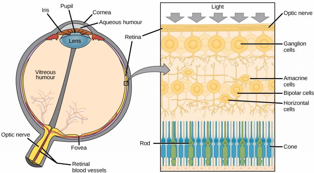
(a) The human eye is shown in cross department. (b) A blowup shows the layers of the retina. Image credit: OpenStax Biology
The vertebrate eye is pretty good at forming squeaky-resolving images, though perhaps we are a piffling biased in making that judgement. Only it turns out that that the vertebrate eye is actually Interahamw from ideal. In point of fact, the cephalopod eye, which looks the Lapplander on the surface but evolved independently, is further better. Here is a compare between the vertebrate and cephalopod eyes:
- Cephalopod eyes move the lense to rive (like in a camera), rather than changing lense shapeas in the craniate eye; the result in vertebrates is long time-related loss of resolution as the lens loses flexibility over time
- Craniate eyes give birth an "inverted" retina, where rakehell vessels and nerves are ahead of the photreceptor cells, unlike in the cephalopod eye where the nerves arebehind the photoreceptor cells; the consequence in vertebrates is the optic disk too age-maternal macular degeneration and increased likelihood of retina detachment (a severe medical emergency) due to the loose association between the retina and the aft of the centre

The vertebrate and class eyes are very similar merely with key differences, including the relative locations of the photoreceptors and sensory neurons (the retina is "inverted" in the vertebrate eye, resulting in a unreasoning spot where the second cranial nerve exits the retina), and the mobile lense in cephalopods compared with a flexible lens in vertebrates. Image credit: modification of work by By Philcha, Public Domain, https://commons.wikimedia.org/w/index.php?curid=4612105
Regardless of the structure of the eye, all photoreception relies on light-absorbing pigment molecules integrated in the photoreceptor cells. This pigment is known as retinal, and it is contained in a protein called opsin . Together, they form a convoluted calledvisual purple, that allow us to find light and color. Both the protein and the pigment are essential for this process:
- The pigment: retinal, reversibly changes shape when it is hit by a photon of light (this process is extremely similar to detection of red vs far-red light by phytochrome in plants.)
- The protein:opsin, holds the retinal pigment and changes shape/activity when the retinal changes shape in reply to concentration of light. Opsins are causative our ability to perceive differences in color or chromaticity. Humans have three color-sensitive opsins: S opsin (short-wavelength opsin), M opsin (culture medium-wavelength opsin), and L opsin (long-wavelength opsin).
- The photoreceptor cells:rods andcones to each one contain a unparalleled opsion that causes the cell the cadre to cost all but sensitive to a specific wavelength of light. Keep in mind that the retinal is ever the same; information technology is the opsin that varies in different photoreceptor cells
- Cones (which are conic) each stop a single case of coloring material-sensitive opsin, making each cone most aware to a fussy hue or color of loose. Though we only have three types of cones (due to the three types of colorise-sensitive opsins), we are competent to detect so much variation in imbue to doh activation of assorted combinations of cones at the same time. Cones require high-levels of light to work, which is why we tail't perceive colouration well in the dark. Cones are intemperately concentrated at thefovea and are useful for focusing on important visual details.
- Rods (which are rod-shaped) each contain a one-fourth type of opsin called rod opsin, which is activated by an intermediate wavelength of twinkle and is capable of working in low-levels of light. Rods are not colour in-sensitive, just allow us to hear in low levels of wanton. Rods are heavily centered at the periphery (outward edges) of the retina, and are useful for detecting movement in our sphere of vision.
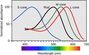
Human rod cells and the different types of cone cells from each one have an optimal wavelength. However, there is considerable overlap in the wavelengths of lightheaded detected. Image credit: OpenStax Biology
This video provides a brief explanation of how we can comprehend so many different colors with only three types of cones:
What happens when a photon activates rhodosin? Unlike all the other sensory systems we have discussed, a pole or retinal conehyperpolarizes when its rhodopsins are activated past light, and itdepolarizes when its rhodopsins are in the inactive. This means that, when in the dark, our rods and cones are depolarized and thus cathartic neurotrasnsmitters to their synapsed emotional disturbance cells. When a rod or cone cell is activated by light, information technology hyperpolarizes andstops emotional neurotransmitter.
Chemoreceptors: Taste (Gustation) and Smell (Olfaction)
The info below was adapted from OpenStax Biota 36.3
Taste, also calledsense of taste, and smell, also namedolfaction, are interconnected senses. Both involve molecules of the stimulus entering the body and soldering to receptors, relying onchemoreceptors, receptors that are sensitive to specific chemicals. Even as each photoreceptor cell is painful to a specific wavelength of light supported on the opsin present in the cell, each chemoreceptor cell is sensitive to a particular molecule supported happening the protein receptor here in the cell.
Taste: the Gustatory System
The primary tastes noticed by humans are sweet, ferment, bitter, salty and umami (savoriness, which tends to suggest that a nutrient is high in protein).
Detecting a taste relies on activating of specific chemical receptors in gustatory sensation receptor cells ( gustatory receptors). When the specific chemical (tastant) binds the receptor, the receptor cell becomes depolarized and releases neurotransmitter happening its synapsed afferent nerve cell. As in new sensorial systems, specificity in tasting occurs because each taste sense organ cell has only one type of protein receptor which is sensitive to either sweet, glum, bitter, salty, or umami. The process of depolarization differs based along the specific type of sense experience sensory receptor:
- A salty tastant (containing NaCl) provides the atomic number 11 ions (Sodium+) that enter the taste receptor cells and excite them directly.
- Turn tastants are acids cause an increase hydrogen ion (H+) concentrations in the taste receptor cells, thus depolarizing them.
- Sweet, bitter, and umami tastants cause activation of an enzyme that causes opening of an ion line, thus depolarizing the taste sensory receptor cells.
- Spiciness isn't heard by taste buds at all, but is actually due to activation of pain receptors (nociceptors)! (More on pain perception at the bottom of this page)
The primary organ of taste is the mouthful bud. Ataste bud is a bundle of sense datum receptors (taste receptor cells) that are located within the bumps on the tongue calledpapillae (remarkable: papilla). For each one predilection bud contains every last five types of sensation receptors (the taste map is totally specious), which are elongated cells with hair-like processes named microvilli at the tips that extend into the taste bud pore. Tastants essential be dissolved in saliva to bind with and stimulate the receptors happening the microvilli, which is why the sense of savour isn't as strong when your mouth is dry.
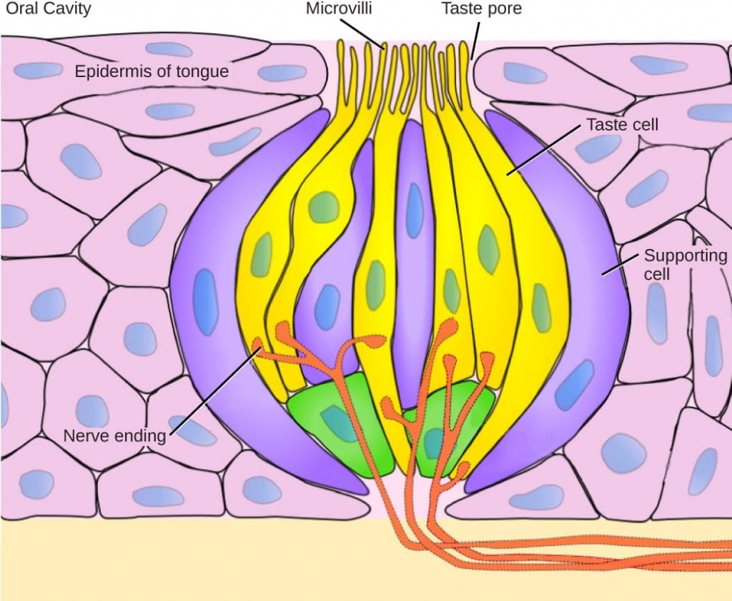
Pores in the tongue allow tastants to interact with and activate gustatory receptors in the taste sensation bud. Each receptor cell contains the receptors for only peerless type of tastant, but each taste bud contains entirely types of taste receptor cells. (credit: OpenStax Biota, modification of work by Vincenzo Rizzo)
Smell: the Olfactory System
Flavor includes a lot more only the 5 primary tastes; most people can detect a difference between the sweet flavors of different types of fruit rather than detecting all of them as just "sweet." But the nuance of flavor doesn't actually come through from gustation the least bit; IT comes from our sense of smell. This is why many populate temporarily recede their gustatory modality when they sustain a cold or other severe nasal congestion.
All odors that we perceive are molecules in the air we breathe. If a substance does not release molecules into the air from its surface, it has no smell. And if a frail or other animal does non accept a receptor that recognizes a specific molecule, and then that molecule has no smell. Human beings have near 350 olfactory sense organ subtypes that work in various combinations to allow us to good sense about 10,000 different odors. (As you may follow expecting by directly, each olfactory receptor cell has only one type of olfactory receptor protein, meaning each receptor cellphone is specific to solitary one character of odorant.) For comparison, mice wealthy person about 1,300 olfactory receptor types and therefore probably horse sense many more odors than get along humans.
How does the sentiency of smell work?
- Odorants(odor molecules) enter the nose and dissolve in the exteroception epithelial tissue, located at the back of the nasal cavity. Theolfactory epithelium is a collection of specialized olfactive receptors in the back of the nasal bone cavity that spans an area about 5 centimetre2 in human race. Similar to tastants, which must be liquid in saliva, an odorant molecule must exist dissolved in mucus ready to be noticed by its sensory receptor.
- Anolfactory receptor, which is a dendrite of a specialized sensory neuron, responds when IT binds certain molecules inhaled from the environment aside sending impulses directly to the olfactory bulb of the brain. Humankind have about 12 million olfactory receptors, distributed among hundreds of different receptor types that respond to different odors. Xii million seems like a large number of receptors, but compare that to past animals: rabbits have most 100 million, most dogs have about 1 billion, and bloodhounds, dogs selectively bred for their olfaction, take in about 4 billion. The general size of the olfactory epithelium also differs between species, with that of bloodhounds, for case, being many times larger than that of humanity.
- Each sensory system neuron has a single dendrite inhumed in the olfactory epithelium, and extending from this dendrite are hair-wish cilia that trap odorant molecules. The sensory receptors on the cilia are proteins, and slim variations in the protein sequences make them sensitive to different odorants.
- Each sense modality sensory nerve cell has only one and only type of receptor protein on its cilia, and the receptors are specialized to notice specific odorants, so for each one olfactory neuron is confident of detecting only a single type of odorant molecule.
- When an odorant binds with a sensory receptor that recognizes it, the sensory neuron related with the receptor is is depolarized and relays carry out potentials to the brain.
- Olfactory stimulation is the only centripetal information that directly reaches the cerebral cortex, whereas past sensations are relayed through the thalamus.
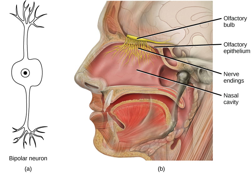
In the human olfactory system, (a) bipolar olfactory neurons extend from (b) the olfactory epithelial tissue, where olfactory receptors are located, to the olfactory bulb. (Image credit: OpenStax Biology, modification of employment by Patrick J. Lynch, medical illustrator; C. Carl Jaffe, MD, heart surgeon)
Our ability to detect and interpret flavor is due to the combination of aesthesis and olfactory senses:
- Taste receptors are causative for the sense of how salty, odoriferous, bitter, sour, or savory a food is, via tastants dissolved in spit
- Olfactory receptors are responsible for the smell of a solid food, via odorants noticed in the olfactory epithelium during chewing, through a process calledretronasal smell(the flow from of air from the spine of the throat up to the olfactory epithelium via the back of the nose out)
This video review how olfaction industrial plant:
Nociceptors: Tissue Damage and Pain
Pain is the name given to nociception, which is the neural processing in response to tissue damage. Pain is caused past both true sources of injury, much As contact with a corrosive chemical, and too by innoxious stimuli that mimic the action of damaging stimuli, such as contact lens with capsaicins, the compounds that cause peppers to taste sizzling (and which are victimised in self-vindication pepper sprays and predestinate local medications). Peppers taste "hot" because the protein receptors that constipate capsaicin open the corresponding Ca channels that are reactive by heat-sensitive thermoreceptors.
There are many different types of nociceptors and we will not describe them therein run, but the important thing to understand is that different types of nociceptors are activated by different types of tissue damage, including extremes of hot or cold, toxic chemicals, and distant mechanical contortion such as stretching, cuts, and crying.
Nociception starts at the sensory receptors, but pain does not actually protrude until it is communicated to the encephalon; hurt is the interpretation of the weave hurt, not the factual input itself. In that location are several nociceptive pathways to and through the brain, including through the thalamus (suchlike about other receptive systems) and straight to the hypothalamus , which modulates the vessel and neuroendocrine functions of the involuntary systema nervosum, where it derriere directly trigger the fight-or-flight response.
This video discusses nociceptor function while explaining how pain relievers sour (the inside information of how pain in the neck relievers work are non relevant to this course, though you English hawthorn find the give-and-take interesting):
Here are much additional videos to help you put this all at once (in a more amusing way than many of the videos above). Distinction that these videos do not allow any new selective information, but they may help you better incorporate all the information previously discussed:
This video provides an attractive review of sharp-eared and balance (bonus points if you set out the motion picture reference!)
This television provides an attractive inspection of craniate eye function:
This television provides an attractive reexamine of sense of smell and gustation:
what specialized neurons detect stimuli like light and sound
Source: https://organismalbio.biosci.gatech.edu/chemical-and-electrical-signals/sensory-systems-i/

Posting Komentar untuk "what specialized neurons detect stimuli like light and sound"